Ricardo Reyes
ACPC Alignment in 3D Slicer
3D Slicer
1
Overview
“AC-PC” alignment is one of many methods for ensuring standardized alignment across neuroimaging. It is mostly applied for MRI imaging and neurosurgical intervention, however, it is an extremely useful strategy for the production of 3D figures using AI segmentation.
3D Slicer’s extension “SlicerNeuro” includes an “ACPC Transform” module. It is recommended to perform realignment prior to AI segmentation because post-alignment may create artificial contours. Generally, the goal of this method is to align the anterior commissure (AC) with the posterior commissure (PC) on an axial plane.
2
‘ACPC Transform’ Module in 3D Slicer
Install the “SlicerNeuro” extension in 3D Slicer (v5.10.0).
AC reference landmarks:
Sagittal: anterior wall of third ventricle
Coronal: positioned between the lateral ventricles (above) and third ventricle (below)
PC reference landmarks:
Sagittal: “C” shaped structure in posterior wall of third ventricle; above midbrain and cerebral aqueduct.
Axial: connects both hemispheres, posterior of third ventricle
Coronal: between the third ventricle (above) and cerebral aqueduct (below)
Defining the midline (on axial):
Superior pontine notch (anterior) where pons and midbrain meet
Anterior falx cerebri
Posterior falx cerebri
Third ventricle
Any additional point in the interhemispheric fissure
3
Applying Transform
In the ‘Data’ module, go to transform hierarchy. Drag the volume into the new transform so the parent is the transform and volume is the child. Right click, harden.
4
Resample the Volume
Go to the module ‘Resample Scalar/Vector/DWI Volume’ and set the input volume as the hardened volume, reference the original volume (pre-hardened), and create new output.
5
Comparing alignment before and after AI segmentation
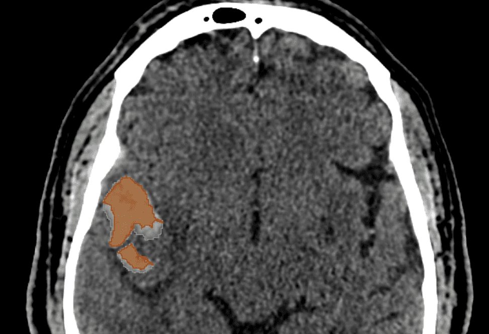
Segmentation Key:
red = post-alignment / green = pre-alignment
Both the red and green segmentations had the same transform except green was applied before AI segmentation with TotalSegmentator. The image shows that there is a real difference on whether you apply before vs after. The red also shows sharp lines in segmentation if ACPC is applied after, which creates an unnatural boundary.
6
Sources
Selvaraj A, Bartels R, Beckmann C, Vinke R, Haak K. ACPC-MAP: A protocol for manually aligning structural T1-weighted Magnetic Resonance Images to the Anterior Commissure – Posterior Commissure plane according to clinical standards. bioRxiv. 2026; 01.12.699070; doi:10.64898/2026.01.12.699070
Fedorov A, Beichel R, Kalpathy-Cramer J, et al. 3D Slicer as an image computing platform for the Quantitative Imaging Network. Magn Reson Imaging. 2012; 30(9):1323-1341; doi:10.1016/j.mri.2012.05.001